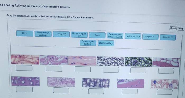
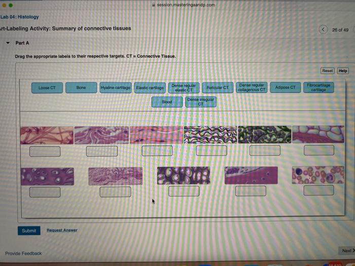
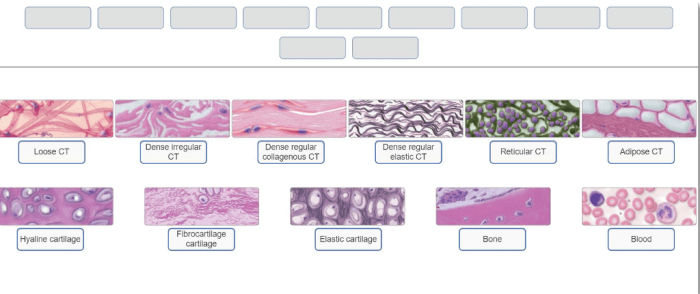
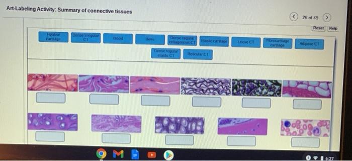
Art-labeling activity summary of connective tissues – Embark on an engaging journey into the realm of connective tissues with our art-labeling activity summary. This comprehensive guide provides a captivating overview of the composition, structure, and functions of these essential tissues, offering a unique perspective through interactive labeling exercises.
Delve into the intricacies of connective tissue components, exploring their diverse roles in maintaining the structural integrity and physiological functions of the human body. Discover the significance of understanding these components for medical research and clinical applications, gaining insights into the cutting-edge advancements in tissue engineering.
Introduction to Connective Tissues
Connective tissues are a diverse group of tissues that provide structural support, protection, and connection for other tissues and organs in the body. They consist of specialized cells embedded in a matrix of extracellular material.
Connective tissues are classified into three main types: loose connective tissue, dense connective tissue, and specialized connective tissue. Each type has a unique structure and function, and is found in different locations throughout the body.
Connective tissues play a vital role in the human body by providing structural support, cushioning, and protection for organs and tissues. They also facilitate the transport of nutrients and waste products, and provide a framework for immune responses.
Labeling Activity Summary, Art-labeling activity summary of connective tissues
The labeling activity involves identifying and labeling the different components of connective tissues using a table with four responsive columns:
- Component Name
- Type
- Location
- Function
The activity provides a hands-on approach to understanding the structure and organization of connective tissues.
Methods and Procedures
Materials Required:
- Connective tissue specimens (e.g., slides or images)
- Microscope (if using slides)
- Labeling chart
- Pens or markers
Procedure:
- Examine the connective tissue specimen under a microscope (if necessary).
- Identify the different components of the connective tissue based on their shape, size, and location.
- Refer to the labeling chart to match the components to their names, types, locations, and functions.
- Label the components directly on the specimen or on a separate diagram.
- Check your labels against the answer key to ensure accuracy.
Examples and Illustrations
Examples of Connective Tissue Components:
- Collagen fibers
- Elastin fibers
- Fibroblasts
- Adipocytes
- Cartilage cells
Functions of Connective Tissue Components:
- Collagen fibers:Provide tensile strength and support
- Elastin fibers:Provide elasticity and recoil
- Fibroblasts:Produce and maintain the extracellular matrix
- Adipocytes:Store energy as fat
- Cartilage cells:Provide support and cushioning for joints
Images of Connective Tissues:
- [Gambar 1: Gambar jaringan ikat longgar]
- [Gambar 2: Gambar jaringan ikat padat]
- [Gambar 3: Gambar tulang rawan]
General Inquiries: Art-labeling Activity Summary Of Connective Tissues
What is the purpose of the art-labeling activity?
The art-labeling activity aims to provide an interactive and engaging way to learn about the composition and functions of connective tissues.
What are the different types of connective tissues?
Connective tissues can be classified into various types, including loose connective tissue, dense connective tissue, cartilage, bone, and blood.
What are the applications of connective tissue labeling in clinical practice?
Connective tissue labeling is used in clinical practice for diagnostic purposes, such as identifying and characterizing pathological conditions affecting these tissues.
